The success of dental implant therapy extends beyond osseointegration to include the quality and quantity of the surrounding soft tissues. While significant breakthroughs have been achieved in hard tissue regeneration, clinicians often find managing soft tissue around dental implants more challenging yet equally crucial.
The peri-implant soft tissue serves as a protective biological seal, contributes to aesthetic outcomes, and plays a vital role in the long-term stability of dental implants. Advances in soft tissue regeneration, such as the use of autografts, allografts, and collagen matrices, are increasingly utilized to enhance tissue volume and improve the esthetics of implant sites, especially in cases of thin or receded gums. Additionally, techniques like the use of growth factors and platelet-rich plasma (PRP) have shown promising results in stimulating tissue regeneration and accelerating healing.
This article explores the latest advancements in soft tissue regeneration dental implant techniques, their benefits, and practical applications in modern implant dentistry.
The Critical Role of Soft Tissue in Dental Implant Success

Fig. 1: Comparison of healthy peri-implant soft tissue (left) versus compromised tissue (right) around dental implants
Understanding the features of the periodontium around natural teeth and peri-implant soft tissue involves recognizing both similarities and differences. The periodontium surrounding natural dentition comprises four key tissues: the periodontal ligament (PDL), cementum, alveolar bone, and gingiva. The gingival tissue consists of the oral epithelium, oral sulcular epithelium, junctional epithelium, and connective tissue attachment.
Dental implants differ from natural teeth in several important ways. There is no periodontal ligament around implants, and the direct structural and functional integration between the implant fixture and the surrounding alveolar bone is defined as osseointegration. Despite this distinction, the soft tissue interface between dental implants and the surrounding gingival tissue shares structural similarities with dentogingival junctions around natural teeth.
Anatomical Differences Between Peri-implant Mucosa and Natural Gingiva
Around natural teeth, gingival fibers run perpendicular to the tooth’s long axis, attaching to and occasionally penetrating the tooth’s structure. Conversely, in implants, these fibers run parallel to the implant’s long axis and do not penetrate its surface. Additionally, while natural teeth boast nine different types of supracrestal fibers enhancing attachment, dental implants typically have only two, potentially resulting in less mechanical resistance in connective tissue adhesion.
When compared to natural dentition, implants exhibit a lower number of blood vessels, relying solely on the large supraperiosteal blood vessel on the outer surface of the alveolar ridge for the peri-implant mucosa’s vascular system. These anatomical differences render dental implants more susceptible to inflammation and subsequent bone loss from microbial challenges, highlighting the importance of proper soft tissue management.
The Peri-implant Phenotype Concept
The peri-implant phenotype encompasses the observable characteristics of the tissues surrounding osseointegrated implants and includes both soft tissue and osseous components. Soft tissue aspects include keratinized tissue width, mucosal thickness, and supracrestal tissue height. These factors significantly influence both the biological and aesthetic outcomes of implant therapy.
Key Components of the Peri-implant Phenotype:
- Keratinized mucosa width (KMW) – The distance from the mucosal margin to the mucogingival junction
- Mucosal thickness (MT) – The horizontal dimension of soft tissue
- Supracrestal tissue height (STH) – The vertical dimension from the bone crest to the mucosal margin
- Peri-implant bone thickness (PBT) – The thickness of bone surrounding the implant
Traditional vs. Modern Soft Tissue Regeneration dental implant Techniques
The field of soft tissue regeneration around dental implants has evolved significantly over the past decades. Understanding both traditional approaches and modern advancements helps clinicians select the most appropriate technique for each clinical scenario.
Traditional Soft Tissue Management Techniques
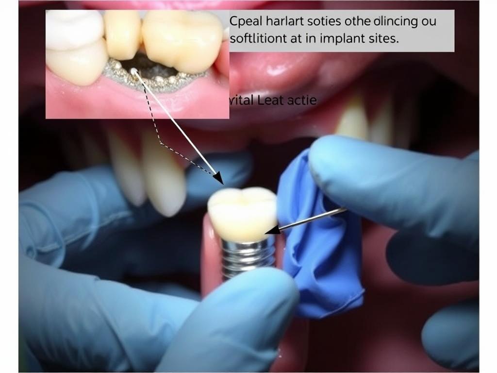
Fig. 2: Traditional autogenous soft tissue grafting procedure around a dental implant
Flap Design Techniques
Early approaches to soft tissue management focused primarily on flap manipulation. Techniques such as the apically positioned flap/vestibuloplasty were designed to extend the depth of the vestibular sulcus, consequently generating keratinized tissue that covers the exposed periosteum or bony surface. Other traditional flap designs include:
- Roll flap technique – Initially developed to correct ridge defects, later modified for use around dental implants
- Palacci technique – Creates papilla-like soft tissue formation between adjacent implants
- Rotational flap designs – Utilizes existing tissue to cover defects without requiring a second surgical site
Autogenous Grafting Procedures
Autogenous grafts have long been considered the gold standard for soft tissue augmentation. These include:
Free Gingival Graft (FGG)
First described by Bjorn in 1963, the free gingival graft involves harvesting epithelialized tissue, typically from the palate, and transplanting it to the recipient site. While effective for increasing keratinized tissue width, FGGs often result in color mismatch and may form scar tissue.
Subepithelial Connective Tissue Graft (SCTG)
Introduced in the 1970s, the SCTG technique involves harvesting connective tissue from beneath the epithelium, usually from the palate. This approach provides excellent esthetic results with better color match and has become the preferred method for soft tissue augmentation in the esthetic zone. Gum Grafting: A Comprehensive Guide to Restoring Your Gum Health
Modern Soft Tissue Regeneration Approaches
Soft Tissue Substitutes
To overcome the limitations of autogenous grafts, such as patient morbidity and limited availability, various soft tissue substitutes have been developed:
| Material Type | Examples | Composition | Clinical Applications | Advantages |
| Acellular Dermal Matrix (ADM) | AlloDerm®, Puros® Dermis | Processed human donor skin with removed cellular components | Increasing soft tissue thickness and keratinized mucosa width | No donor site morbidity, unlimited availability, maintains extracellular matrix structure |
| Xenogeneic Collagen Matrix | Mucograft® | Non-cross-linked porcine bilayer collagen matrix | Increasing keratinized tissue width | Shorter surgical time, lower post-operative pain, better color match |
| Volume-Stable Collagen Matrix | Fibro-Gide® | Porcine, porous, resorbable and volume-stable collagen matrix | Soft tissue volume augmentation | Volume stability, promotes angiogenesis and fibroblast ingrowth |
Growth Factor-Enhanced Regeneration
The application of growth factors represents a significant advancement in soft tissue regeneration. These bioactive molecules can enhance healing and tissue formation:
- Platelet-Rich Fibrin (PRF) – Autologous blood concentrate containing platelets, leukocytes, and growth factors that promote tissue healing and regeneration
- Platelet-Rich Plasma (PRP) – Concentrated platelets in plasma containing various growth factors that stimulate cell proliferation and differentiation
- Recombinant Human Platelet-Derived Growth Factor (rhPDGF) – Specifically promotes fibroblast proliferation and angiogenesis
3D Bioprinting and Tissue Engineering
Emerging technologies in tissue engineering offer promising solutions for soft tissue regeneration:

Fig. 4: 3D bioprinting technology for creating customized soft tissue scaffolds
3D bioprinting allows for the creation of custom-designed scaffolds seeded with patient-derived cells. This approach enables the fabrication of tissue constructs with precise architectural features that mimic natural soft tissue. While still largely experimental in dental applications, early research shows promising results for creating functional soft tissue replacements with vascular networks and cellular organization similar to native tissues.
Enhance Your Clinical Outcomes with Advanced Techniques
Download our comprehensive guide on soft tissue regeneration techniques to improve your implant success rates and achieve predictable aesthetic results.Download Clinical Guide
Benefits of Soft Tissue Regeneration in Implant Dentistry
Proper management of peri-implant soft tissues offers numerous advantages that contribute to both the functional and aesthetic success of dental implant therapy.
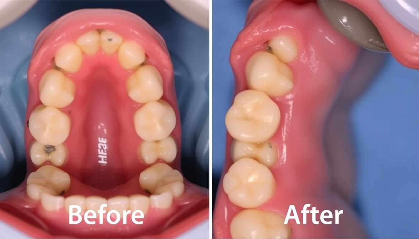
Fig. 5: Before (left) and after (right) soft tissue regeneration around dental implants in the aesthetic zone
Improved Healing and Integration
Adequate soft tissue volume and quality create an optimal environment for implant integration. Research has demonstrated that proper soft tissue management can:
- Enhance wound healing through better vascularization
- Reduce inflammatory responses during the healing phase
- Create a stable biological seal around the implant-abutment interface
- Minimize microbial colonization during the critical healing period
Enhanced Aesthetics
In the aesthetic zone, soft tissue management is particularly crucial for achieving natural-looking results:
- Creates natural soft tissue contours and emergence profiles
- Establishes proper gingival zenith positions and papilla formation
- Masks the underlying implant components and restorative materials
- Prevents mucosal discoloration by providing adequate tissue thickness
- Maintains long-term stability of the gingival margin position
Reduced Infection Risk
A healthy band of keratinized mucosa around dental implants serves as a protective barrier:
- Creates a tight seal against bacterial invasion
- Reduces plaque accumulation and facilitates oral hygiene
- Decreases the incidence of peri-implant mucositis and peri-implantitis
- Minimizes inflammatory responses to bacterial challenges
- Provides resistance to mechanical trauma during function and oral hygiene
Long-term Stability and Maintenance
Recent systematic reviews have highlighted the importance of adequate soft tissue quality and quantity for the long-term success of dental implants. Studies have shown that:
Implants surrounded by an adequate width of keratinized mucosa (≥2 mm) demonstrate significantly less marginal bone loss over time compared to those with insufficient keratinized tissue. A meta-analysis by Thoma et al. (2018) found that soft tissue grafting procedures resulted in a significant reduction in marginal bone loss and bleeding on probing over long-term follow-up periods.
Additionally, adequate mucosal thickness (≥2 mm) has been associated with better preservation of crestal bone levels. This is particularly important in the aesthetic zone, where even minor changes in tissue dimensions can compromise the visual outcome. Proper soft tissue management also facilitates professional maintenance and patient home care, contributing to the overall longevity of the implant restoration.
4.7
Overall Impact on Implant Success
Aesthetic Improvement
4.8
Peri-implant Health
4.7
Long-term Stability
4.5
Patient Satisfaction
4.6
Challenges and Solutions in Soft Tissue Regeneration
Despite significant advancements, soft tissue regeneration around dental implants presents several challenges that clinicians must address to achieve optimal outcomes.
- Limited availability of autogenous donor tissue
- Patient morbidity associated with harvesting procedures
- Unpredictable shrinkage of grafted tissues
- Biocompatibility concerns with substitute materials
- Technical sensitivity of surgical procedures
- Cost considerations for advanced materials
- Anatomical limitations in certain clinical scenarios
- Development of minimally invasive harvesting techniques
- Utilization of soft tissue substitutes to reduce donor site morbidity
- Overcorrection strategies to compensate for tissue shrinkage
- Advanced biocompatible materials with improved integration
- Specialized training and microsurgical approaches
- Cost-effective treatment planning and material selection
- Customized approaches based on individual patient anatomy
Biocompatibility Considerations

Fig. 6: Histological comparison of tissue responses to different soft tissue regeneration materials
The biocompatibility of materials used for soft tissue regeneration is a critical factor in treatment success. Autogenous tissues remain the gold standard due to their excellent biocompatibility and integration potential. However, when using substitute materials, clinicians must consider:
- Host immune response to allogeneic and xenogeneic materials
- Degradation patterns and timing of substitute materials
- Integration with surrounding tissues and vascularization potential
- Long-term stability and tissue maintenance
Recent advancements in material processing techniques have significantly improved the biocompatibility of substitute materials. For example, modern acellular dermal matrices undergo sophisticated decellularization processes that effectively remove immunogenic components while preserving the extracellular matrix structure essential for tissue integration.
Cost-Effectiveness Analysis
The economic aspect of soft tissue regeneration procedures is an important consideration in clinical decision-making. While advanced materials and techniques may have higher initial costs, they often provide better long-term outcomes with fewer complications.
| Technique/Material | Initial Cost | Procedure Time | Patient Morbidity | Long-term Value |
| Autogenous SCTG | Low | Longer (requires harvest) | Higher | Excellent |
| Acellular Dermal Matrix | Moderate-High | Shorter | Lower | Good |
| Xenogeneic Collagen Matrix | Moderate-High | Shorter | Lower | Good |
| PRF-Enhanced Procedures | Low-Moderate | Moderate | Lower | Good |
A comprehensive cost-effectiveness analysis should consider not only the immediate procedural costs but also the long-term benefits, including reduced complication rates, decreased need for revision surgeries, and improved patient satisfaction. In many cases, investing in appropriate soft tissue management early in the treatment process proves more cost-effective than addressing complications later.
Clinical Case Studies
The following case studies illustrate the application of soft tissue regeneration techniques in different clinical scenarios and their impact on treatment outcomes.
Case 1: Soft Tissue Management in the Aesthetic Zone

Fig. 7: Case 1 – Soft tissue management in the aesthetic zone using connective tissue grafting
Patient Profile and Initial Situation
A 32-year-old female patient presented with a missing maxillary central incisor (tooth #8) following traumatic injury. Clinical examination revealed adequate bone volume for implant placement but significant soft tissue deficiency on the buccal aspect. The patient had high aesthetic expectations and was concerned about the final appearance of the restoration.
Treatment Approach
A staged approach was implemented:
- Initial soft tissue augmentation using a subepithelial connective tissue graft harvested from the palate
- Implant placement after 8 weeks of soft tissue healing
- Second-stage surgery with additional soft tissue modification using a roll flap technique
- Provisional restoration to develop optimal emergence profile
- Final ceramic restoration after tissue maturation
Outcome and Discussion
The final result demonstrated excellent soft tissue contours with natural emergence profile and papilla formation. The staged approach allowed for predictable tissue augmentation and precise control of the aesthetic outcome. Key factors contributing to success included:
- Addressing soft tissue deficiency before implant placement
- Using autogenous connective tissue for optimal integration and tissue quality
- Careful provisional restoration design to guide tissue maturation
- Allowing adequate healing time between surgical phases
At the 2-year follow-up, the peri-implant tissues remained stable with no signs of recession or inflammation, demonstrating the long-term benefits of comprehensive soft tissue management.
Case 2: Management of Peri-implant Soft Tissue Deficiency in the Posterior Region
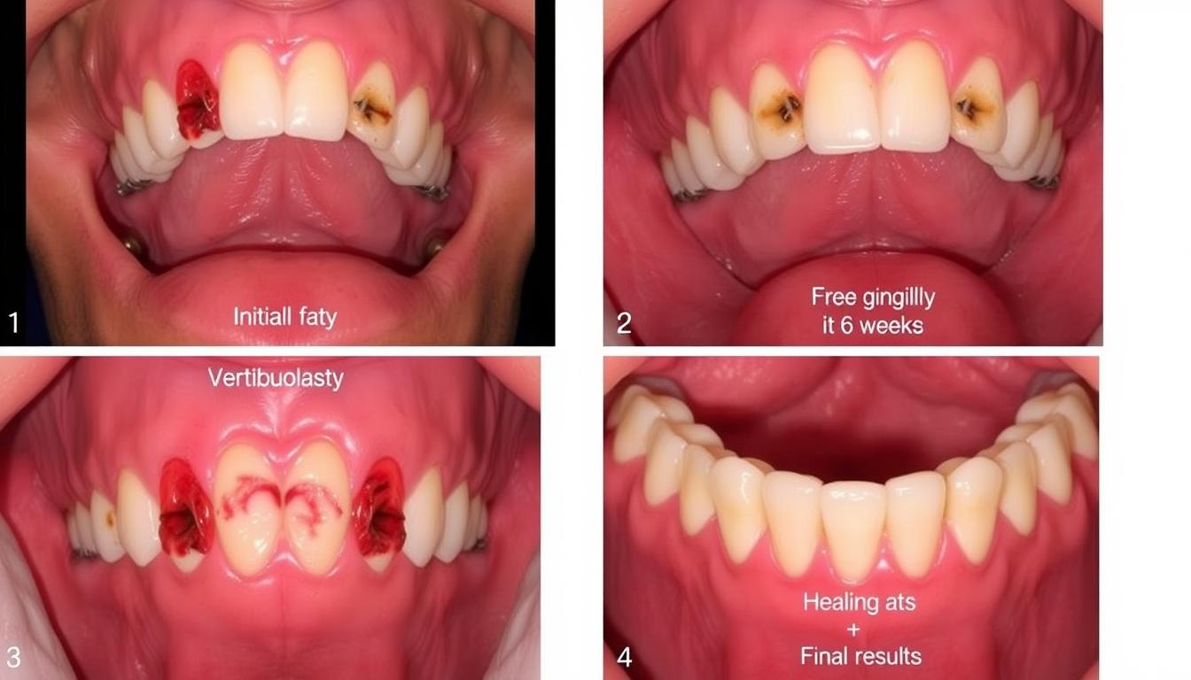
Fig. 8: Case 2 – Management of peri-implant soft tissue deficiency in the posterior region
Patient Profile and Initial Situation
A 58-year-old male patient presented with two dental implants in the mandibular posterior region (teeth #19 and #20) that had been restored 3 years prior. The patient reported discomfort during oral hygiene procedures and occasional bleeding around the implants. Clinical examination revealed a complete lack of keratinized tissue around both implants, with signs of mucositis and early bone loss on radiographic evaluation.
Treatment Approach
The treatment focused on establishing an adequate band of keratinized tissue:
- Removal of the existing prosthesis to access the peri-implant tissues
- Vestibuloplasty procedure with apically positioned flap
- Placement of a xenogeneic collagen matrix (Mucograft®) to increase keratinized tissue width
- Modification of the prosthetic components to enhance tissue adaptation
- Reinstallation of the modified prosthesis after 6 weeks of healing
Outcome and Discussion
The intervention resulted in a significant increase in keratinized tissue width (from 0 mm to 3.5 mm) around both implants. At the 1-year follow-up, the patient reported complete resolution of discomfort during oral hygiene procedures, and clinical examination showed healthy peri-implant tissues with no bleeding on probing. Radiographic evaluation demonstrated stabilization of bone levels.
This case illustrates the importance of keratinized tissue around dental implants, particularly in the posterior regions where oral hygiene access is more challenging. The use of a xenogeneic collagen matrix provided an effective alternative to autogenous grafting, reducing patient morbidity while achieving the desired clinical outcome.
Improve Your Soft Tissue Management Skills
Access our collection of detailed case studies demonstrating various soft tissue regeneration techniques and their long-term outcomes.Download Case Collection
Timing Considerations for Soft Tissue Regeneration
The timing of soft tissue interventions plays a crucial role in the success and predictability of regenerative procedures. Different approaches can be implemented at various stages of implant therapy.
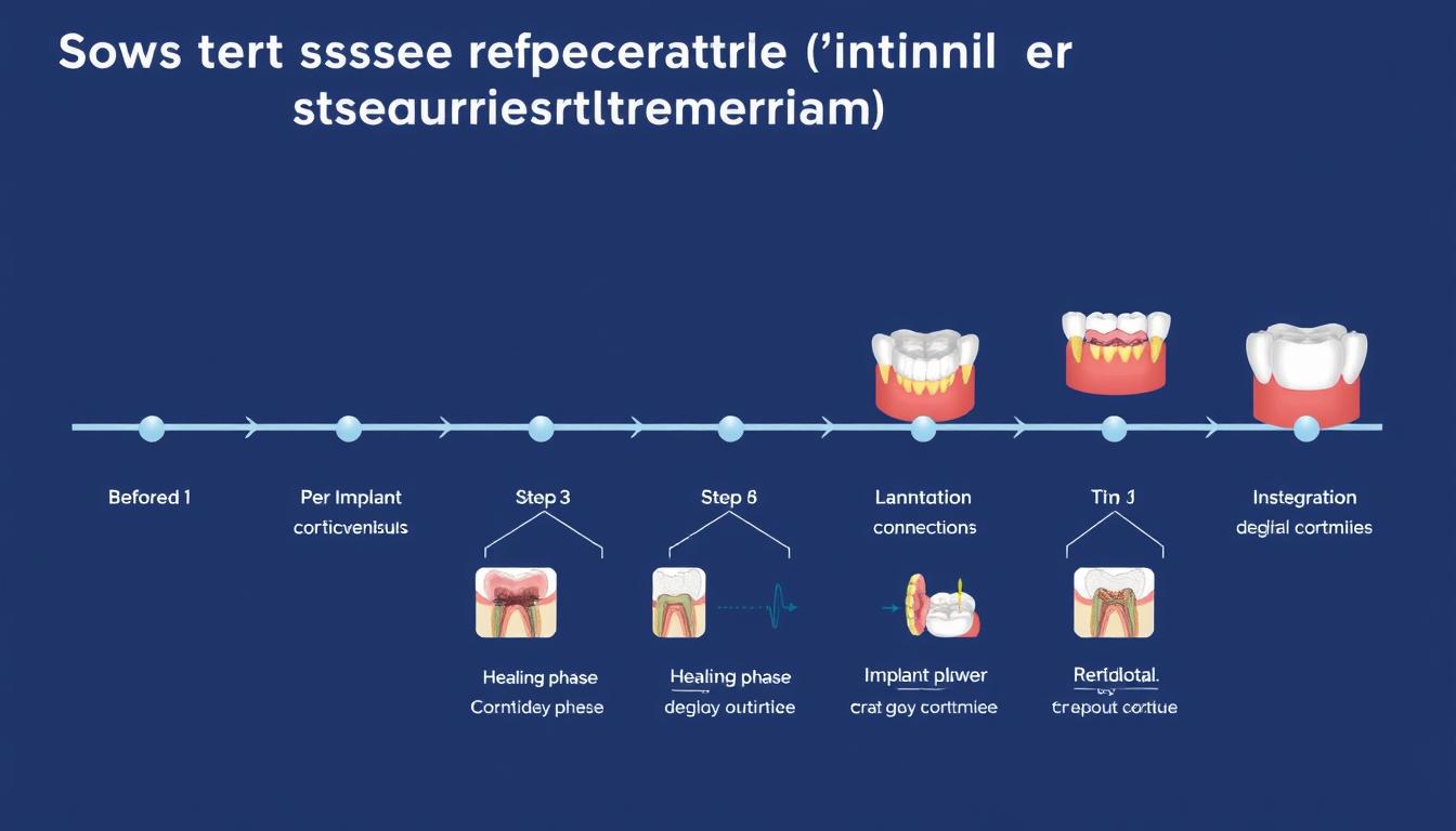
Fig. 9: Timeline showing optimal stages for soft tissue regeneration interventions in implant therapy
| Timing | Technique | Indications | Advantages | Limitations |
| Before Implant Placement | Soft tissue augmentation with SCTG or substitutes | Significant soft tissue deficiency, thin biotype | Improves tissue quality for subsequent procedures, highly predictable | Extends treatment time, additional surgery |
| During Implant Placement | Simultaneous soft tissue grafting with implant surgery | Immediate implants, moderate tissue deficiency | Reduces number of surgeries, addresses tissue needs early | More complex procedure, potential for complications |
| During Healing Phase | Soft tissue augmentation before second-stage surgery | Developing soft tissue deficiencies, preparation for uncovery | Allows focused soft tissue management, good healing potential | Additional surgery, potential impact on implant healing |
| At Abutment Connection | Soft tissue modification during second-stage surgery | Minor soft tissue deficiencies, keratinized tissue enhancement | Combines with planned second-stage surgery, shapes emergence profile | Limited potential for major augmentation |
| After Restoration | Corrective soft tissue procedures | Developing recession, aesthetic concerns | Addresses specific deficiencies | Less predictable, may require prosthesis removal |
Research suggests that early intervention for soft tissue management yields more predictable results. A systematic review by Thoma et al. (2014) concluded that soft tissue augmentation procedures performed before or during implant placement demonstrated superior outcomes compared to later interventions. This is particularly relevant in the aesthetic zone, where tissue dimensions significantly impact the final appearance of the restoration.
Frequently Asked Questions About Soft Tissue Regeneration
What is the minimum width of keratinized tissue needed around dental implants?
While there is ongoing debate about the minimum required width, current evidence suggests that at least 2 mm of keratinized mucosa is beneficial for maintaining peri-implant health. However, this requirement may vary based on several factors including implant position, restoration type, and patient-specific factors such as oral hygiene capability and tissue biotype. In the posterior regions and in patients with compromised oral hygiene, a wider band of keratinized tissue (3-4 mm) may be advantageous.
Are soft tissue substitutes as effective as autogenous grafts?
Current evidence indicates that while autogenous grafts remain the gold standard, certain soft tissue substitutes can provide comparable results in specific clinical scenarios. For increasing keratinized tissue width, xenogeneic collagen matrices have shown similar outcomes to free gingival grafts with reduced patient morbidity. For volume augmentation, autogenous connective tissue grafts still demonstrate superior results, although volume-stable collagen matrices are showing promising outcomes in recent studies. The selection between autogenous grafts and substitutes should be based on the specific clinical situation, patient preferences, and the clinician’s experience.
How does soft tissue thickness affect implant aesthetics?
Soft tissue thickness significantly impacts the aesthetic outcome of implant restorations in several ways. A minimum thickness of 2 mm is generally recommended to mask the underlying implant components and prevent grayish discoloration of the mucosa, particularly when using titanium abutments. Thicker tissues also provide better support for maintaining proper emergence profiles and gingival contours. Additionally, adequate tissue thickness contributes to the stability of the gingival margin position, reducing the risk of recession over time. In cases with thin tissue biotypes, soft tissue augmentation and/or the use of ceramic abutments should be considered to optimize aesthetic outcomes.
Can soft tissue deficiencies be corrected after the final restoration is placed?
While it is possible to address soft tissue deficiencies after the final restoration is placed, these interventions are generally less predictable and more technically challenging than preventive approaches. Corrective procedures may include connective tissue grafting for recession coverage or free gingival grafts for keratinized tissue augmentation. In some cases, the restoration may need to be removed temporarily to facilitate proper tissue management. The success of these interventions depends on various factors including the severity of the deficiency, the position of the implant, the design of the restoration, and the patient’s healing capacity. Whenever possible, soft tissue management should be addressed proactively before the final restoration is delivered.
What role does the emergence profile play in soft tissue stability?
The emergence profile of the implant restoration significantly influences soft tissue stability and aesthetics. An appropriately designed emergence profile provides proper support for the surrounding tissues while facilitating oral hygiene access. The critical contour (area immediately apical to the gingival margin) determines the position and shape of the gingival zenith, while the subcritical contour (extending from the critical contour to the implant platform) affects tissue thickness and support. Overcontoured restorations can cause pressure-induced recession, while undercontoured designs may lead to food impaction and inadequate tissue support. Provisional restorations play a crucial role in gradually developing the optimal emergence profile before the final restoration is fabricated.
Future Trends in Soft Tissue Regeneration
Fig. 10: Advanced stem cell research for next-generation soft tissue regeneration
The field of soft tissue regeneration continues to evolve rapidly, with several promising developments on the horizon that may revolutionize clinical practice in the coming years.
Stem Cell-Based Approaches
Stem cell therapy represents one of the most promising frontiers in soft tissue regeneration. Current research focuses on:
- Harvesting and expanding gingival mesenchymal stem cells for targeted tissue regeneration
- Developing cell-seeded scaffolds that can be customized to specific defect geometries
- Creating “off-the-shelf” cell-based products that eliminate the need for autogenous tissue harvesting
- Utilizing induced pluripotent stem cells (iPSCs) to generate patient-specific gingival tissues
Early clinical trials have demonstrated the safety and potential efficacy of stem cell-based approaches, particularly for challenging cases with significant tissue deficiencies.
Advanced Biomaterials and Delivery Systems

Fig. 11: Next-generation biomaterials with controlled release of bioactive factors
The development of novel biomaterials with enhanced biological properties is advancing rapidly:
Smart Biomaterials
Responsive materials that can adapt to the local tissue environment, releasing bioactive factors in response to specific stimuli such as pH changes, enzyme activity, or mechanical forces. These materials can provide more targeted and efficient delivery of growth factors and other therapeutic agents.
Nanofibrous Scaffolds
Electrospun scaffolds with nanoscale architecture that mimics the natural extracellular matrix, providing optimal conditions for cell attachment, proliferation, and differentiation. These scaffolds can be engineered with specific mechanical properties and degradation rates to match the requirements of soft tissue regeneration.
Digital Technologies and Personalized Approaches
Digital technologies are increasingly being integrated into soft tissue management workflows:
- 3D digital planning for precise soft tissue augmentation based on the desired final outcome
- Computer-aided design and manufacturing (CAD/CAM) of customized scaffolds tailored to individual defect morphology
- Intraoral scanning and digital monitoring of soft tissue changes over time
- Artificial intelligence algorithms for predicting treatment outcomes and optimizing surgical approaches
These technologies enable more predictable and personalized approaches to soft tissue regeneration, potentially improving both clinical efficiency and patient outcomes.
“The future of soft tissue regeneration lies at the intersection of biology, materials science, and digital technology. By combining our understanding of tissue healing with advanced biomaterials and precise digital workflows, we can develop more predictable and less invasive approaches to creating healthy peri-implant tissues.”
– Dr. Daniel S. Thoma, University of Zurich
Conclusion
Soft tissue regeneration has emerged as a critical component of successful implant therapy, influencing both biological and aesthetic outcomes. The peri-implant soft tissues serve as a protective barrier, contribute to aesthetic appearance, and play a vital role in long-term implant health and stability.
Modern approaches to soft tissue management offer clinicians a variety of options, from traditional autogenous grafting techniques to advanced biomaterials and growth factor-enhanced procedures. The selection of appropriate techniques should be based on a thorough assessment of the individual clinical situation, patient-specific factors, and desired outcomes.
Timing considerations are crucial in soft tissue management, with early intervention generally providing more predictable results. A proactive approach to addressing soft tissue deficiencies before they become problematic is recommended whenever possible.
As research continues to advance, the integration of stem cell technologies, smart biomaterials, and digital workflows promises to further enhance the predictability and efficiency of soft tissue regeneration procedures. These developments will likely lead to less invasive approaches with reduced patient morbidity and improved outcomes.
By understanding and implementing current best practices in soft tissue regeneration while staying informed about emerging technologies, clinicians can optimize the functional and aesthetic results of implant therapy, ultimately enhancing patient satisfaction and quality of life.
Stay Updated with the Latest Advancements
Subscribe to our newsletter for regular updates on soft tissue regeneration techniques, clinical cases, and research developments.Subscribe Now
References
- Thoma DS, Naenni N, Figuero E, et al. Effects of soft tissue augmentation procedures on peri-implant health or disease: A systematic review and meta-analysis. Clin Oral Implants Res. 2018;29(Suppl 15):32-49. doi: 10.1111/clr.13114
- Avila-Ortiz G, Gonzalez-Martin O, Couso-Queiruga E, Wang HL. The peri-implant phenotype. J Periodontol. 2020;91(3):283-288. doi: 10.1002/JPER.19-0566
- Thoma DS, Buranawat B, Hämmerle CH, Held U, Jung RE. Efficacy of soft tissue augmentation around dental implants and in partially edentulous areas: a systematic review. J Clin Periodontol. 2014;41(Suppl 15):S77-S91. doi: 10.1111/jcpe.12220
- Zeltner M, Jung RE, Hämmerle CH, Hüsler J, Thoma DS. Randomized controlled clinical study comparing a volume-stable collagen matrix to autogenous connective tissue grafts for soft tissue augmentation at implant sites: linear volumetric soft tissue changes up to 3 months. J Clin Periodontol. 2017;44:446-453. doi: 10.1111/jcpe.12697
- Sculean A, Gruber R, Bosshardt DD. Soft tissue wound healing around teeth and dental implants. J Clin Periodontol. 2014;41(Suppl 15):S6-22. doi: 10.1111/jcpe.12206