The rehabilitation of atrophic posterior maxilla presents significant challenges for dental implantologists. Limited bone height, poor bone quality, and proximity to the maxillary sinus often complicate traditional implant placement. Pterygoid implants offer a compelling alternative to conventional bone grafting procedures, sinus lifts, and zygomatic implants. This article explores the anatomical considerations, surgical techniques, clinical applications, and recent advancements in pterygoid implant technology, providing dental professionals with comprehensive insights into this valuable treatment option.
Overview of Pterygoid Implants

Figure 1: Anatomical position of a pterygoid implant passing through the maxillary tuberosity into the pterygoid process
Anatomical Location and Considerations
Pterygoid implants are characterized by their unique anatomical pathway. These implants originate in the tuberosity region of the maxilla and follow an oblique mesiocranial direction, proceeding posteriorly toward the pyramidal process of the palatine bone. They subsequently extend upward between both wings of the pterygoid process of the sphenoid bone.
The implant traverses what some authors describe as a “corridor” comprising:
- The maxillary tuberosity
- The pyramidal process of the palatine bone
- The pterygoid process of the sphenoid bone
- Terminating at the pterygoid fossa
Unlike the posterior maxillary alveolar ridge, the pterygoid process is not a tooth-dependent structure and remains relatively stable regardless of tooth pathology or sinus pneumatization. This makes it a reliable anchor point for implant placement in patients with significant posterior maxillary atrophy.
Unique Features
Several characteristics distinguish pterygoid implants from conventional dental implants:
- Length: Typically between 13-20mm to achieve adequate engagement with the pterygoid plates
- Angulation: Placed at a 45-60° angle relative to the occlusal plane
- Cortical Engagement: Anchors into the dense cortical bone at the junction of the palatine and pterygoid processes
- Graftless Solution: Eliminates the need for sinus augmentation or extensive bone grafting
Enhance Your Implant Expertise
Looking to incorporate pterygoid implants into your practice? Our team offers personalized training and mentorship for dental professionals.Schedule a Consultation
Comparison with Alternative Techniques

Figure 2: Comparison between pterygoid implants, zygomatic implants, and traditional implants with sinus lift
| Feature | Pterygoid Implants | Zygomatic Implants | Traditional Implants with Sinus Lift |
| Surgical Complexity | Moderate to High | High | Moderate |
| Treatment Time | Single-stage possible | Single-stage possible | Multiple stages (3-9 months) |
| Bone Grafting Required | No | No | Yes |
| Patient Morbidity | Low to Moderate | Moderate to High | Moderate |
| Anesthesia | Local | General often preferred | Local |
| Immediate Loading Potential | High | High | Low |
| Prosthetic Considerations | Angled abutments required | Complex prosthetic design | Standard prosthetics |
Advantages Over Traditional Approaches
Compared to conventional sinus augmentation procedures, pterygoid implants offer several distinct advantages:
Reduced Treatment Time
Pterygoid implants eliminate the healing period associated with bone grafting, potentially reducing overall treatment time by 3-6 months. This allows for faster restoration of function and aesthetics for patients with posterior maxillary atrophy.
Decreased Surgical Morbidity
By avoiding sinus augmentation procedures, pterygoid implants reduce the risk of complications such as membrane perforation, graft infection, and sinusitis. The technique also eliminates donor site morbidity associated with autogenous bone harvesting.
Comparison with Zygomatic Implants
While both pterygoid and zygomatic implants provide graftless solutions for the atrophic maxilla, they differ in several important aspects:
- Pterygoid implants typically require only local anesthesia, whereas zygomatic implants often necessitate general anesthesia
- Zygomatic implants involve a more complex anatomical pathway with proximity to critical structures like the orbit and infraorbital nerve
- Pterygoid implants generally result in less patient discomfort and faster recovery compared to zygomatic implants
- Zygomatic implants may be preferred in cases of extreme anterior maxillary atrophy, while pterygoid implants excel in posterior rehabilitation
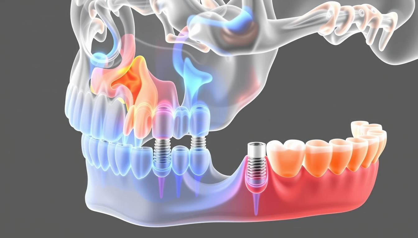
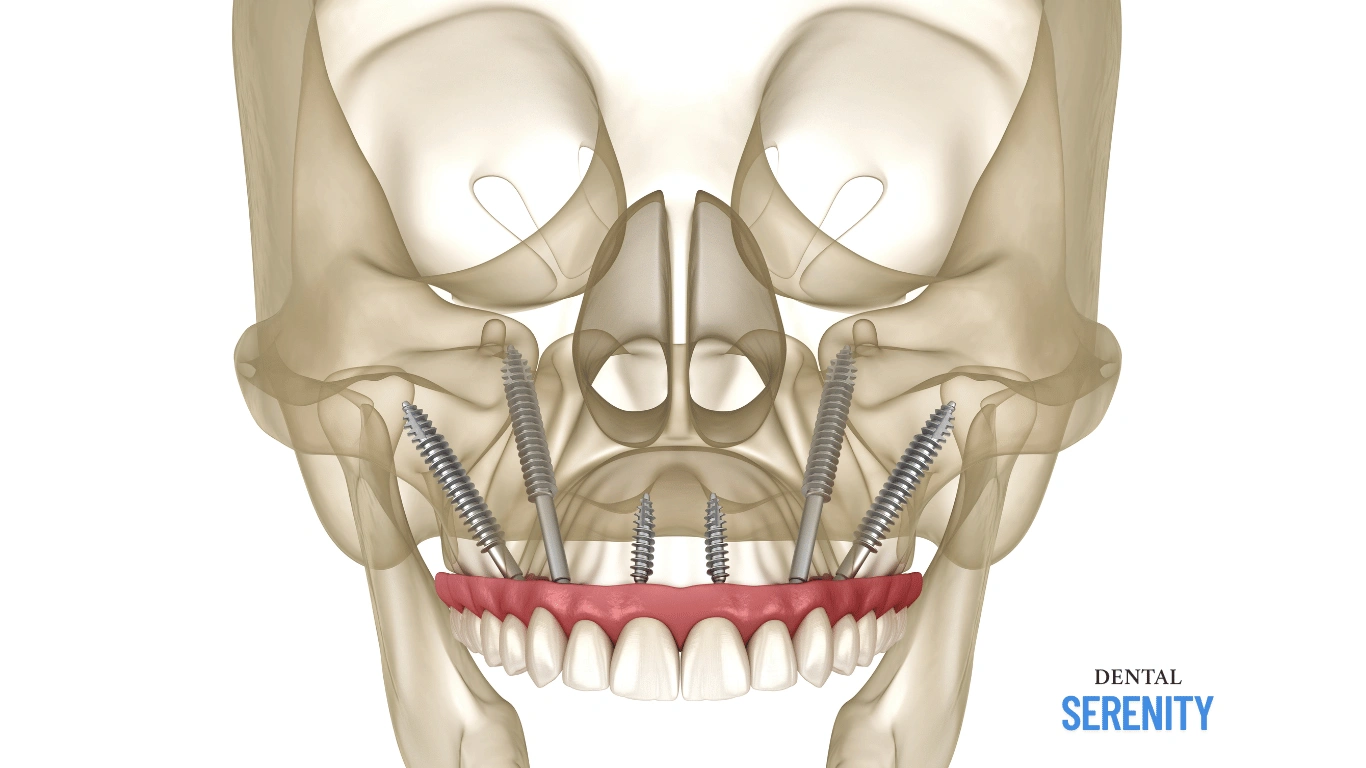
Figure 3: 3D reconstruction showing optimal placement of pterygoid implants from a prosthetic-driven perspective
Surgical Techniques and Success Rates
Surgical Protocol
The placement of pterygoid implants requires precise technique and thorough understanding of the complex anatomy. The standard surgical approach follows these key steps:
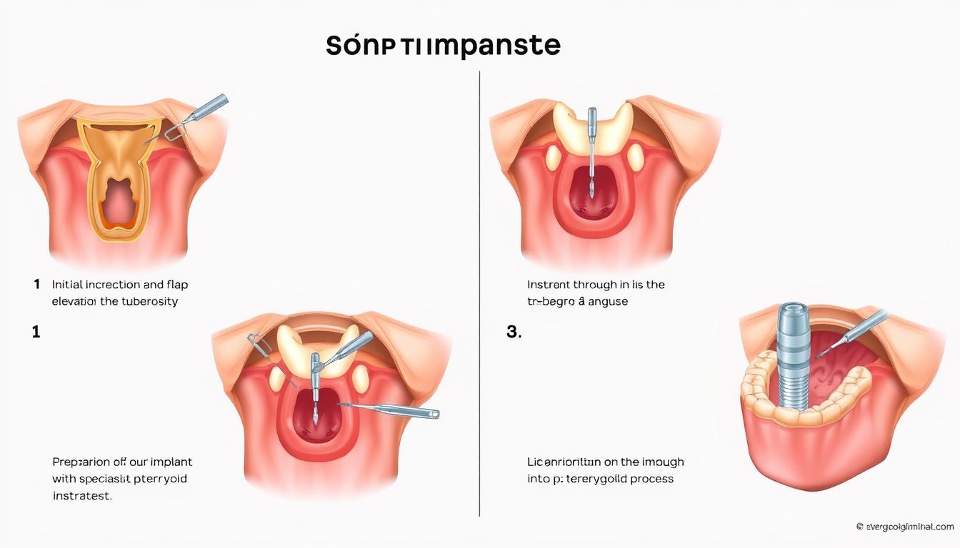
Figure 4: Step-by-step surgical technique for pterygoid implant placement
- Incision and Exposure: A midcrestal incision is made in the pterygomaxillary region with a vertical releasing incision mesial to the homolateral canine. A full-thickness mucoperiosteal buccal flap is elevated to expose the surgical site.
- Site Preparation: Drilling begins at the second molar region with an angulation of 45-55° relative to the occlusal plane. The drill path proceeds posteriorly and superiorly toward the pterygoid plates.
- Implant Selection: Implants of appropriate length (typically 15-18mm) and diameter (4-4.5mm) are selected to ensure engagement with the pterygoid process.
- Implant Placement: The implant is inserted following the prepared path, with the apical portion engaging the cortical bone of the pterygoid plates.
- Closure: The flap is repositioned and sutured, with the implant platform positioned for subsequent prosthetic connection.
Success Rates and Long-term Outcomes
Multiple clinical studies have demonstrated high success rates for pterygoid implants, comparable to or exceeding those of conventional implants in the posterior maxilla:
98.3%
Average Success Rate
7+ Year Success Rate
97.6%
5-7 Year Success Rate
98.2%
3-5 Year Success Rate
100%
A recent meta-analysis by Araujo et al. (2019) reported a cumulative success rate of 94.87% for pterygoid implants, while Fernández-Valerón et al. (2020) documented a 96.45% success rate in their long-term study. These findings suggest that pterygoid implants offer predictable and durable outcomes for posterior maxillary rehabilitation.
Master the Pterygoid Implant Technique
Access our comprehensive surgical protocol with detailed illustrations and video demonstrations.Download Complete Surgical Guide
Clinical Applications
Atrophic Maxilla Cases
Pterygoid implants are particularly valuable in cases of severe posterior maxillary atrophy, where conventional implant placement would require extensive bone augmentation. The technique is indicated in the following scenarios:
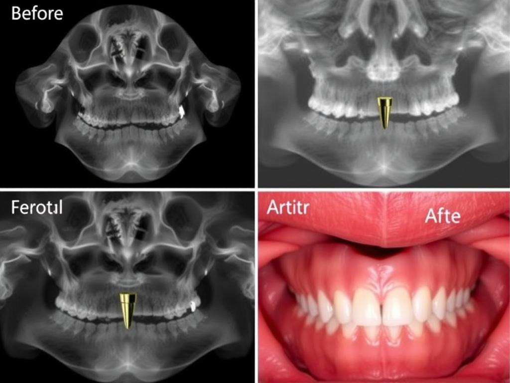
Figure 5: Clinical case showing pterygoid implant placement in a patient with severe maxillary atrophy
- Residual bone height less than 4mm in the posterior maxilla due to sinus pneumatization
- Severe horizontal and vertical resorption of the alveolar ridge
- Failed previous bone grafting procedures
- Patients with contraindications to extensive bone augmentation
- Cases requiring immediate or early loading protocols
Full-Arch Rehabilitation
In full-arch rehabilitation of the edentulous maxilla, pterygoid implants provide valuable posterior support, eliminating the need for distal cantilevers and improving biomechanical stability:
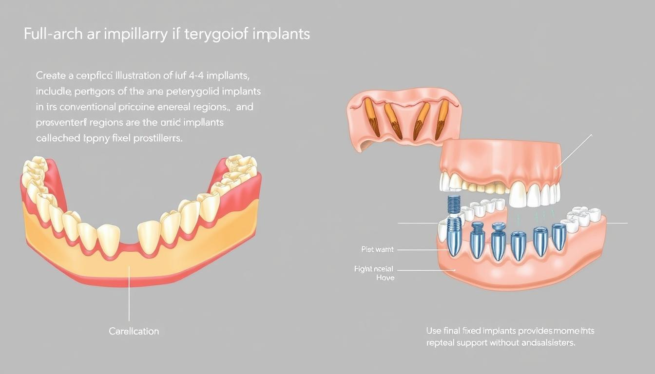
Figure 6: Full-arch rehabilitation using pterygoid implants in combination with anterior implants
All-on-4/6 Concept
Pterygoid implants can be integrated into the All-on-4 or All-on-6 concept, replacing tilted posterior implants. This approach maximizes the anterior-posterior spread of implants, creating a more favorable distribution of occlusal forces and reducing stress on individual implants.
Immediate Loading Protocols
ZYGOMATIC IMPLANTSThe dense cortical bone engagement achieved with pterygoid implants often results in excellent primary stability, facilitating immediate loading protocols. This allows for the delivery of fixed provisional prostheses on the day of surgery, significantly improving patient satisfaction.
Combination with Other Implant Types
Pterygoid implants can be effectively combined with conventional, zygomatic, or short implants to create comprehensive solutions for complex maxillary rehabilitation:

Figure 7: Hybrid approach combining pterygoid implants with conventional and zygomatic implants
- Conventional implants in the anterior maxilla with pterygoid implants posteriorly
- Unilateral zygomatic implant with contralateral pterygoid implant
- Short implants in areas with moderate bone height combined with pterygoid implants
- Strategic implant placement based on available bone and prosthetic requirements
Benefits and Potential Complications
Benefits
- Eliminates need for sinus augmentation and bone grafting
- Reduces overall treatment time by 3-6 months
- Decreases surgical morbidity and patient discomfort
- Allows for immediate or early loading protocols
- Provides excellent biomechanical support for posterior maxilla
- Eliminates distal cantilevers in full-arch prostheses
- High long-term success rates (94-98%)
- Lower economic cost compared to multiple surgical interventions
Potential Complications
- Technically demanding surgical procedure
- Risk of hemorrhage from pterygoid venous plexus
- Potential injury to greater palatine artery
- Limited access and visibility during surgery
- Prosthetic challenges due to implant angulation
- Risk of postoperative trismus
- Potential for paresthesia (rare)
- Steep learning curve for clinicians
Managing Potential Complications
While pterygoid implants are generally associated with minimal complications, proper management strategies are essential:

Figure 8: Anatomical illustration highlighting critical structures to avoid during pterygoid implant placement
| Potential Complication | Prevention Strategy | Management Approach |
| Hemorrhage | Thorough preoperative CBCT analysis; careful surgical technique | Local hemostatic agents; pressure application; consider embolization for severe cases |
| Inadequate Primary Stability | Proper implant selection; engagement with pterygoid plates | Consider larger diameter or longer implant; delayed loading protocol |
| Trismus | Careful soft tissue management; avoid pterygoid muscle trauma | Anti-inflammatory medication; physical therapy; muscle relaxants |
| Prosthetic Complications | Proper implant angulation; use of angled abutments | Custom abutments; CAD/CAM frameworks; screw-retained prostheses |
| Paresthesia | Knowledge of neurovascular anatomy; careful technique | Observation; neurotrophic medications; rarely surgical exploration |
Recent Advancements in Materials and Design
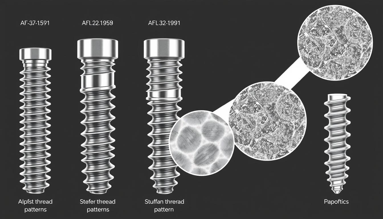
Figure 9: Modern pterygoid implant designs showing advancements in thread patterns and surface treatments
Implant Design Innovations
Recent years have seen significant advancements in pterygoid implant design, enhancing their performance and ease of use:
- Tapered Designs: Modern pterygoid implants feature tapered designs that facilitate insertion and improve initial stability in the pterygoid region
- Progressive Thread Patterns: Advanced thread geometries provide enhanced engagement with the cortical bone of the pterygoid plates
- Platform Switching: Incorporation of platform switching concepts to preserve crestal bone and improve long-term stability
- Specialized Drilling Protocols: Development of site-specific drilling sequences optimized for the unique anatomy of the pterygomaxillary region
Surface Technology Advancements
Surface modifications have significantly improved the osseointegration potential of pterygoid implants:
Hydrophilic Surfaces
Modern implant surfaces with enhanced hydrophilicity accelerate the initial healing phase, promoting faster osseointegration in the challenging pterygoid region. These surfaces demonstrate improved protein adsorption and cellular attachment, critical factors for success in areas with limited bone quality.
Nanotopography
Implants with nanoscale surface features have shown enhanced osteoblast activity and bone-to-implant contact. This is particularly beneficial for pterygoid implants, where maximizing bone-implant interface is essential for long-term stability and function.
Digital Planning and Guided Surgery
The integration of digital technologies has revolutionized pterygoid implant placement:
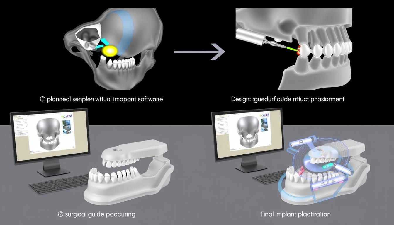
Figure 10: Digital planning and guided surgery for pterygoid implant placement
- CBCT-Based Planning: High-resolution CBCT imaging allows for detailed analysis of the pterygomaxillary anatomy and precise virtual implant positioning
- Custom Surgical Guides: CAD/CAM technology enables the fabrication of patient-specific surgical guides for accurate pterygoid implant placement
- Dynamic Navigation: Real-time surgical navigation systems provide intraoperative guidance, enhancing precision and safety
- Prosthetically Driven Planning: Digital workflows ensure that implant positioning is optimized for subsequent prosthetic rehabilitation
Stay at the Forefront of Implant Dentistry
Connect with specialists and access the latest research and techniques in advanced implantology.Join Our Professional Network
Frequently Asked Questions

Figure 11: Pterygoid implant placement showing key anatomical landmarks and surgical access
What is the ideal length and diameter for pterygoid implants?
The ideal dimensions for pterygoid implants typically range from 15-18mm in length and 4.0-4.5mm in diameter. These dimensions ensure adequate engagement with the pterygoid plates while providing sufficient strength to withstand occlusal forces. However, implant selection should be customized based on individual patient anatomy as determined by preoperative CBCT analysis. Recent studies suggest that implants with a minimum length of 13mm can achieve successful outcomes when properly positioned to engage the cortical bone of the pterygoid process.
What is the learning curve for pterygoid implant placement?
The learning curve for pterygoid implant placement is relatively steep due to the complex anatomy and limited visibility of the pterygomaxillary region. Clinicians should undergo specialized training and preferably complete 10-15 procedures under mentorship before performing the technique independently. Proficiency typically requires 25-30 cases. The use of digital planning and guided surgery can significantly reduce the learning curve and enhance predictability for clinicians new to the technique. Hands-on cadaver courses are highly recommended as part of the training process.
Can pterygoid implants be immediately loaded?
Yes, pterygoid implants can be immediately loaded when adequate primary stability is achieved (typically insertion torque >35 Ncm). The dense cortical bone engagement in the pterygoid region often provides excellent initial stability, making these implants suitable candidates for immediate loading protocols. Studies by Balshi et al. and Peñarrocha et al. have demonstrated high success rates with immediately loaded pterygoid implants in full-arch rehabilitations. However, the decision for immediate loading should be made on a case-by-case basis, considering factors such as bone quality, implant stability, occlusal scheme, and patient-specific risk factors.
How do pterygoid implants affect prosthetic design?
Pterygoid implants are typically placed at a 45-60° angle relative to the occlusal plane, which necessitates specific prosthetic considerations. Angled abutments (17-30°) are commonly required to achieve prosthetic parallelism. CAD/CAM technology has significantly improved the precision of prosthetic frameworks for pterygoid implants. Screw-retained prostheses are generally preferred over cement-retained options to facilitate maintenance and retrievability. The distal position of pterygoid implants eliminates the need for cantilevers, allowing for a more favorable distribution of occlusal forces throughout the prosthesis.
What are the contraindications for pterygoid implants?
Contraindications for pterygoid implants include severe trismus or limited mouth opening (10 cigarettes/day), severe bruxism without appropriate management, and inadequate pterygoid process volume as determined by CBCT analysis. A thorough preoperative assessment is essential to identify potential contraindications and mitigate risks.
Conclusion
Pterygoid implants represent a valuable addition to the implantologist’s armamentarium for addressing the challenges of posterior maxillary atrophy. By leveraging the stable bone of the pterygoid process, these implants provide a graftless solution that reduces treatment time, decreases patient morbidity, and delivers predictable long-term outcomes. The high success rates documented in the literature, combined with recent advancements in implant design, surface technology, and digital workflows, have established pterygoid implants as a scientifically validated and predictable treatment option.
While the technique demands specialized training and meticulous attention to anatomical considerations, the benefits for appropriate cases are substantial. As implant dentistry continues to evolve toward minimally invasive approaches and accelerated treatment protocols, pterygoid implants will likely play an increasingly important role in the comprehensive management of the atrophic maxilla.
1 thought on “Pterygoid Implants: Advanced Solutions for Posterior Maxillary Rehabilitation”