Modern dentistry has evolved significantly with advanced imaging technologies that provide detailed views of oral structures. Among these innovations, Cone Beam Computed Tomography (CBCT) has revolutionized dental diagnostics and treatment planning. This three-dimensional imaging technique offers unprecedented detail of teeth, bone, nerve pathways, and soft tissues in a single scan, allowing dental professionals to provide more accurate diagnoses and precise treatment plans. The integration of CBCT scan dental technology enhances the ability to evaluate complex cases, such as implant placement, root canal treatments, and orthodontic assessments, by giving a comprehensive view of the patient’s dental anatomy. With this advanced tool, dentists can plan treatments with higher precision, improving patient outcomes and reducing the risk of complications.
What is a CBCT Scan dental and How Does It Work?

A dental CBCT scanner uses a cone-shaped X-ray beam to create detailed 3D images
Dental cone beam computed tomography (CBCT) is a specialized type of X-ray equipment that generates three-dimensional images of dental structures, soft tissues, nerve pathways, and bone in the craniofacial region. Unlike conventional CT scanners, CBCT uses a cone-shaped X-ray beam that rotates around the patient, capturing multiple images from different angles.
During a CBCT scan, the machine’s arm rotates 360 degrees around the patient’s head, collecting between 150 to 200 high-resolution two-dimensional images. These images are then digitally combined to create a detailed 3D model that can be viewed from any angle and in different planes (axial, coronal, and sagittal).
The technology behind CBCT differs from traditional dental X-rays in several important ways:
Traditional Dental X-rays
- Produce 2D images with structure overlap
- Limited view of complex anatomy
- Multiple exposures often needed
- Fixed perspective
- Lower radiation dose per image
CBCT Scans
- Generate detailed 3D images
- Show complex relationships between structures
- Single scan captures entire region
- Images can be viewed from multiple angles
- Higher radiation dose than single X-ray
CBCT technology has become increasingly accessible in dental practices due to its compact size, relatively lower cost compared to medical CT scanners, and the valuable diagnostic information it provides. TRIOS Intraoral Scanners: Revolutionizing Digital Dentistry
Clinical Applications of CBCT Scans in Dentistry
CBCT imaging has transformed numerous areas of dental practice by providing detailed visualization of oral and maxillofacial structures. Here are the key clinical applications where CBCT scans have proven invaluable:
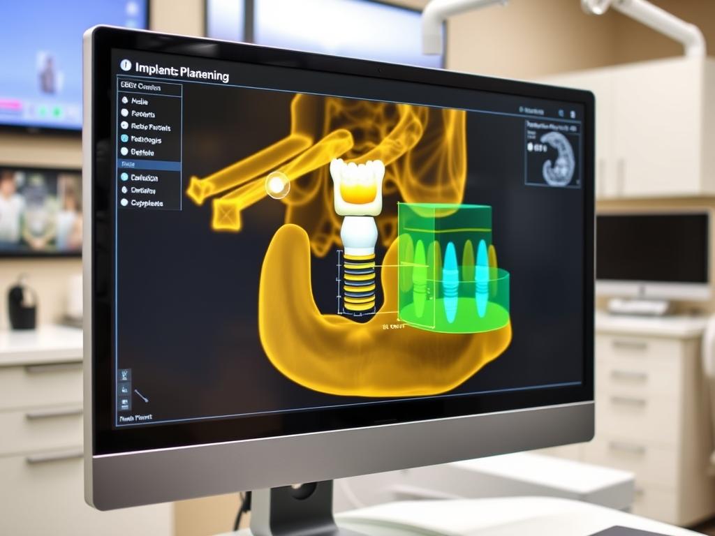
CBCT scan used for precise dental implant planning and placement
1. Dental Implant Planning and Placement
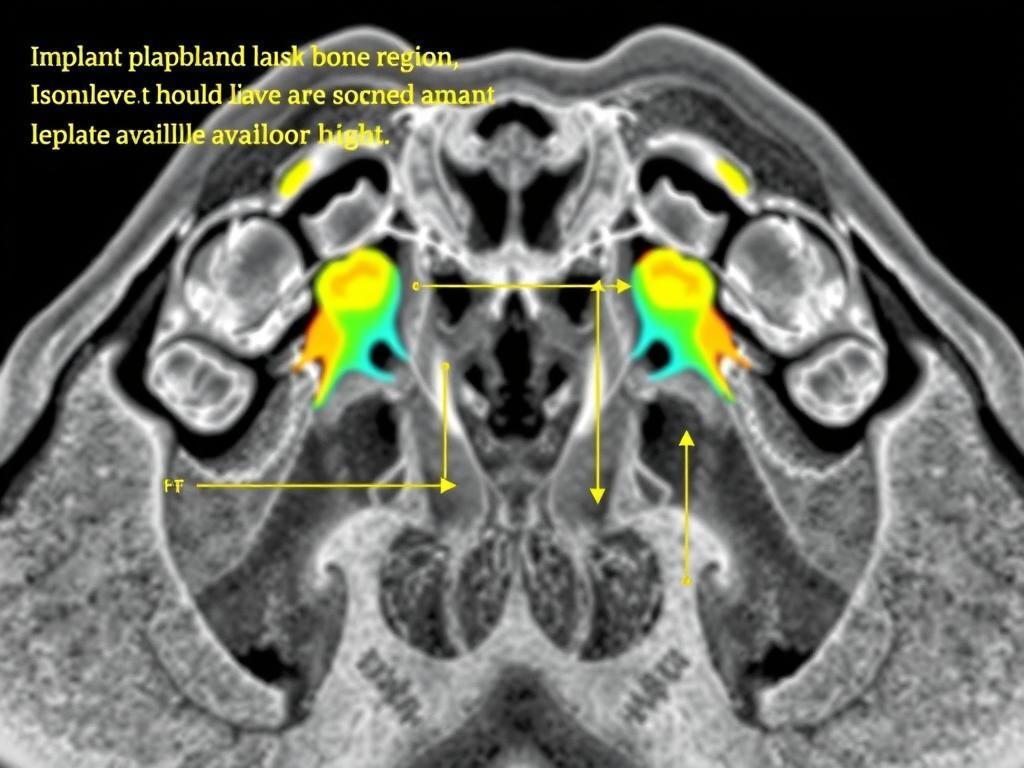
CBCT scans provide critical information for successful implant procedures, including:
- Precise measurement of bone height, width, and density
- Visualization of vital anatomical structures (nerves, sinuses)
- Virtual implant placement before surgery
- Creation of surgical guides for accurate implant positioning
- Post-surgical assessment of implant integration
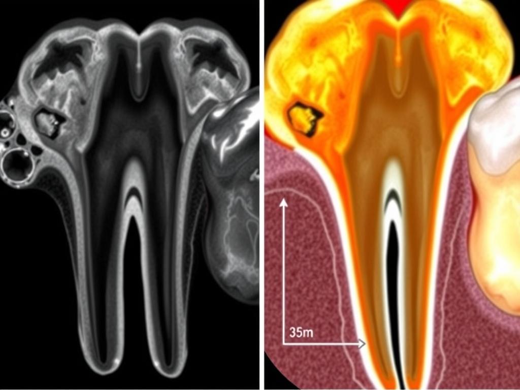
2. Endodontic (Root Canal) Diagnosis and Treatment
CBCT enhances endodontic care by revealing:
- Number and morphology of root canals
- Presence and extent of periapical lesions
- Root fractures that may not be visible on 2D X-rays
- Internal and external root resorption
- Relationship between roots and surrounding structures
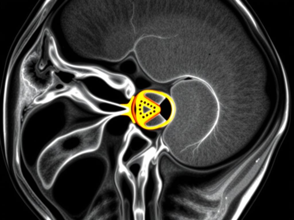
Detailed CBCT visualization of temporomandibular joint anatomy
3. TMJ (Temporomandibular Joint) Analysis
For patients with TMJ disorders, CBCT provides:
- Visualization of joint space and condylar position
- Detection of bone changes and abnormalities
- Assessment of joint function and morphology
- Evaluation of the effects of TMJ disorders on surrounding structures
4. Orthodontic Assessment and Planning
Orthodontists use CBCT for:
- Evaluation of tooth position and inclination
- Assessment of bone thickness for safe tooth movement
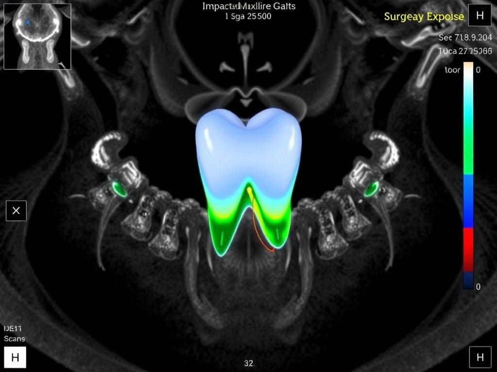
- Visualization of impacted teeth and their relationship to other structures
- Airway analysis for sleep apnea concerns
- Planning for orthognathic surgery
5. Oral Surgery and Pathology
CBCT assists surgeons by showing:
- Precise location and extent of cysts, tumors, and other pathologies
- Relationship between impacted teeth and vital structures
- Fracture patterns in facial trauma
- Anatomical variations that may affect surgical approaches
CBCT visualization of periodontal bone loss and furcation involvement
6. Periodontal Assessment
For periodontal (gum) disease evaluation, CBCT reveals:
- Three-dimensional view of bone loss patterns
- Furcation involvements (where bone loss occurs between roots)
- Dehiscence and fenestration defects
- Assessment of regenerative periodontal treatment outcomes
7. Airway Analysis
CBCT is valuable for examining:
- Upper airway dimensions and volume
- Nasal passage and sinus evaluation
- Contributing factors to sleep-disordered breathing
- Effects of orthodontic or orthognathic treatment on airway
CBCT vs. Traditional Dental X-rays: A Comparison
Understanding the differences between CBCT and conventional dental X-rays helps patients and practitioners make informed decisions about which imaging method is most appropriate for specific clinical situations.
| Feature | Traditional Dental X-rays | CBCT Scans |
| Image Dimension | 2D (flat images) | 3D (volumetric data) |
| Radiation Dose | Lower (5-10 μSv for a single periapical) | Higher (30-500 μSv depending on FOV) |
| Detail Resolution | Limited by overlapping structures | High detail with submillimeter resolution |
| Cost | $20-150 depending on type and number | $150-750 depending on region scanned |
| Scan Time | Seconds per image | 5-40 seconds for complete scan |
| Anatomical Coverage | Limited to specific areas | Entire dental arch or full craniofacial region |
| Soft Tissue Visualization | Poor | Limited but better than 2D X-rays |
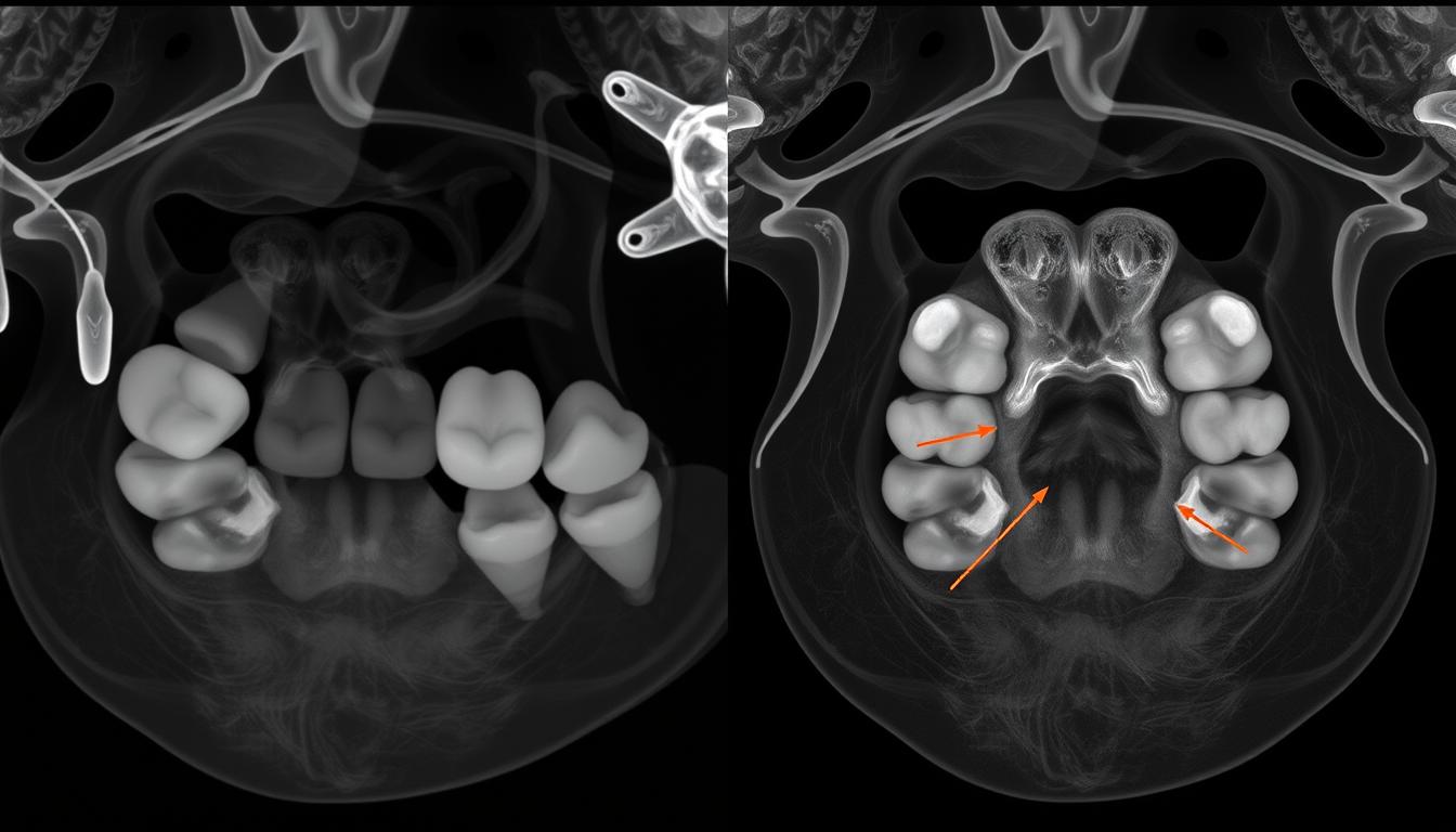
Comparison between traditional 2D X-ray (left) and CBCT scan (right) of the same dental region
Radiation Considerations
While CBCT scans deliver more radiation than conventional dental X-rays, the dose is significantly lower than medical CT scans. The effective radiation dose from a CBCT scan ranges from 29-477 μSv, depending on the field of view (FOV) and the specific CBCT unit used.
For perspective, a CBCT scan delivers radiation equivalent to approximately 3-48 days of natural background radiation. By comparison, a full-mouth series of traditional dental X-rays delivers about 35-150 μSv, while a medical CT of the head can deliver 2,000 μSv or more.
Modern CBCT units allow for field-of-view limitation, which restricts the X-ray beam to the area of interest, reducing unnecessary radiation exposure. Additionally, using personal protection (thyroid collar) can further reduce radiation dose by up to 40%. ICAD Scanners in Dental Implantology: Precision, Workflow, and Clinical Advantages
What to Expect During a CBCT Scan
If your dentist has recommended a CBCT scan, knowing what to expect can help ease any concerns about the procedure. The process is straightforward, painless, and typically takes less than 30 minutes from start to finish.

Patient positioned in a dental CBCT scanner for imaging
Before the Scan: Preparation
- Little to no special preparation is required for a CBCT scan
- You’ll be asked to remove metal objects that might interfere with imaging, such as jewelry, eyeglasses, hearing aids, and removable dental appliances
- Inform your dentist if you are pregnant or think you might be pregnant
- Wear comfortable clothing without metal zippers or buttons in the neck area
During the Scan: Procedure
- You’ll either sit in an upright chair or lie down on an examination table, depending on the type of CBCT scanner
- The technician will position your head using guides to ensure it remains still during the scan
- You may be provided with a bite block to help maintain the correct position
- The scanner’s arm will rotate around your head in a complete or partial 360-degree rotation
- The scan itself typically takes between 10-40 seconds
- You’ll be asked to remain very still and may be instructed to hold your breath briefly to prevent movement
- The procedure is completely painless – you won’t feel the X-rays
After the Scan: Results
- You can resume normal activities immediately after the scan
- The images are processed by computer software and made available to your dentist
- Your dentist will analyze the images and discuss the findings with you
- The results may be available immediately or at a follow-up appointment, depending on your dentist’s protocol
Safety Tip: While CBCT scans use ionizing radiation, the benefits of an accurate diagnosis generally outweigh the minimal risks involved when the scan is medically necessary. Always discuss any concerns about radiation exposure with your dental professional.
Case Studies: How CBCT Changed Treatment Outcomes
The following case studies illustrate how CBCT imaging has significantly improved diagnosis and treatment planning in real clinical scenarios. These examples demonstrate the practical benefits of this advanced imaging technology.

Case 1: Hidden Root Fracture Detection
Clinic: Precision Dental Imaging Center
Scenario: A 45-year-old patient presented with persistent pain in an endodontically treated molar. Traditional X-rays showed no apparent issues.
CBCT Finding: The scan revealed a vertical root fracture that was not visible on conventional radiographs.
Outcome: Instead of attempting retreatment of the root canal, which would have failed, the tooth was extracted and replaced with an implant, resolving the patient’s pain and preventing further complications.
Case 2: Complex Impacted Canine
Clinic: Advanced Orthodontic Solutions
Scenario: A 14-year-old orthodontic patient had an impacted maxillary canine. Conventional X-rays suggested possible root resorption of adjacent teeth.
CBCT Finding: The scan provided precise localization of the impacted tooth and showed it was positioned palatally with minimal contact with adjacent roots.
Outcome: The orthodontist and oral surgeon were able to plan a conservative surgical approach and successful orthodontic traction, preserving all adjacent teeth that might otherwise have been extracted based on 2D imaging alone.
Case 3: Unexpected Anatomical Variation
Clinic: Comprehensive Implant Institute
Scenario: A 62-year-old patient needed an implant in the upper molar region. Initial panoramic X-rays suggested adequate bone height.
CBCT Finding: The scan revealed an unexpected anatomical variation with the maxillary sinus extending much lower than anticipated, leaving insufficient bone for standard implant placement.
Outcome: The treatment plan was modified to include a sinus lift procedure before implant placement. This prevented potential sinus perforation and implant failure that might have occurred if the procedure had been planned using only 2D images.
These cases highlight how CBCT imaging provides critical information that can significantly alter treatment approaches, leading to better outcomes, fewer complications, and more predictable results for patients.
Frequently Asked Questions About CBCT Scans

Dental professional reviewing CBCT scan results with a patient
Are CBCT scans safe for children?
CBCT scans should be used judiciously in children since they are more sensitive to radiation. However, when medically necessary, the benefits of an accurate diagnosis typically outweigh the minimal risks. Dental professionals follow the ALARA principle (As Low As Reasonably Achievable) when determining radiation exposure, especially for pediatric patients. Modern CBCT units allow for field-of-view limitation and reduced settings for children. The scan should only be performed when the diagnostic information cannot be obtained through conventional, lower-dose imaging methods.
How much does a dental CBCT scan cost?
The cost of a dental CBCT scan typically ranges from $150 to $750, depending on the region being scanned, the facility, and your geographic location. Some dental insurance plans may cover part of the cost if the scan is deemed medically necessary. However, coverage varies widely between providers and plans. It’s advisable to check with your insurance company before undergoing the procedure. Many dental offices offer payment plans or financing options for patients without insurance coverage.
How does the radiation from a CBCT scan compare to other X-rays?
A CBCT scan delivers more radiation than traditional dental X-rays but significantly less than a medical CT scan. For comparison:
- Single digital periapical X-ray: 5-10 μSv
- Digital panoramic X-ray: 14-24 μSv
- Full-mouth series of digital X-rays: 35-150 μSv
- CBCT scan (depending on field of view): 30-500 μSv
- Medical CT of the head: 2,000+ μSv
For perspective, humans receive about 3,000 μSv annually from natural background radiation sources.
Will my insurance cover a CBCT scan?
Insurance coverage for CBCT scans varies widely. Some dental and medical insurance plans will cover these scans when they’re deemed medically necessary for diagnosis or treatment planning. Coverage may be more likely for complex cases such as implant planning, impacted teeth, or suspected pathology. To determine coverage, provide your insurance company with the specific procedure code (usually CDT code D0364-D0368 depending on the scan type) and ask about your benefits. Your dental office may also be able to help with insurance pre-authorization.
How long does it take to get CBCT scan results?
The actual CBCT scanning process takes only 10-40 seconds. The images are then processed by computer software, which typically takes just a few minutes. In many cases, your dentist can review the results with you during the same appointment. However, for complex cases requiring detailed analysis or consultation with specialists, it may take a few days before your dentist discusses the findings with you. The timeframe for receiving results should be clarified with your dental provider before the procedure.
Does a CBCT scan hurt?
No, a CBCT scan is completely painless. The procedure is non-invasive and doesn’t involve any injections, incisions, or physical contact with the scanning equipment. You’ll simply need to remain still for a short period (typically less than a minute) while the scanner rotates around your head. Some patients may experience mild discomfort from having to maintain a specific position or from using a bite block to stabilize their head, but the scan itself causes no pain or sensation.
Conclusion: The Future of Dental Imaging

Modern dental practice with integrated CBCT and digital workflow systems
CBCT scanning represents a significant advancement in dental imaging technology, providing unprecedented detail and diagnostic capabilities that were previously unavailable in the dental office. As the technology continues to evolve, we can expect even more refined applications and integration with other digital dental technologies.
While CBCT scans deliver higher radiation doses than conventional dental X-rays, the diagnostic benefits often outweigh the minimal risks when the scan is medically necessary. Dental professionals carefully consider the ALARA principle (As Low As Reasonably Achievable) when recommending imaging procedures, especially for pediatric patients.
For patients, CBCT imaging translates to more accurate diagnoses, more predictable treatment outcomes, fewer complications, and often less invasive procedures. The ability to visualize oral structures in three dimensions has transformed many aspects of dental care, from implant placement to endodontic therapy to orthodontic treatment planning.
If your dental professional recommends a CBCT scan, you can feel confident that this advanced imaging technology will provide valuable information that can significantly improve your treatment experience and outcome. Always discuss any concerns about radiation exposure or the necessity of the scan with your dental provider, who can explain the specific benefits for your individual case.
Want to Learn More About Advanced Dental Imaging?
Discover how modern imaging technologies are improving dental diagnosis and treatment outcomes.Explore Dental Imaging Guide
Wondering if a CBCT Scan Could Help Your Dental Condition?
Speak with a dental professional to determine if CBCT imaging is appropriate for your specific needs.Find a CBCT Specialist Near You
Preparing for Your CBCT Scan?
Download our free patient preparation guide to ensure you’re ready for your upcoming CBCT scan appointment.Download Preparation Guide
Have a Complex Dental Case?
Our specialists can evaluate your situation and determine if advanced imaging like CBCT could help provide better diagnosis and treatment planning.Request a Case Evaluation
Have More Questions About CBCT Scans?
Our dental professionals are ready to answer your specific questions about CBCT imaging and how it might benefit your oral health.Ask Your Question
kssp1i
8r2c2x